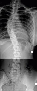
The x-ray is used for assessment and subsequent documentation of spinal deformity. The x-ray should reveal the entire spine, and should be made standing. X-rays in the recumbent position are not necessary as they do not provide much information. The roentgenographic series of the spine in an a/p perspective should consist of a thoracic part and a lumbar part in order to minimize the distorsion of the picture by dispersion of the rays. However, one x-ray film may be large enough for smaller children. An x-ray from the lateral perspective is also very useful. It is important that the scope of the x-ray is wide enough in oder to be able to see the ribs and the pelvis.
An x-ray is associated with radiation, therefore it is not advisable to take repeated pictures if this is not absolutely necessary. On the other hand, it would not be wise to avoid taking a repeated x-ray when the risk of scoliosis progression is high. The amount of radiation received from modern x-ray apparatus is slight and less harmful than making the wrong treatment decision due to lack of information.
In comparison, the amount of radiation received when flying from Europe to America – an amount usually not deemed harmful -is equivalent to the radiation received from taking three x-rays.
Medical professionals have differing opinions about how often repeat x-rays should be taken. In the period when scoliosis progression is high, x-rays can be repeated every 3 months. However, when the deformity has stabilized, once a year is enough.
The degree of curvature seen on the x-ray is calculated using the Cobb method (see figure). The correct measurement of the Cobb angle entails determining the end vertebrae of each curve and drawing lines along the top endplate of the upper end vertebra and bottom endplate of the lower end vertebra. T he place where the lines meet forms the Cobb angle. Should the lines meet outside the picture, perpendicular lines may be drawn for easier calculation.


